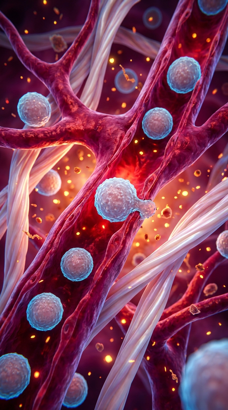
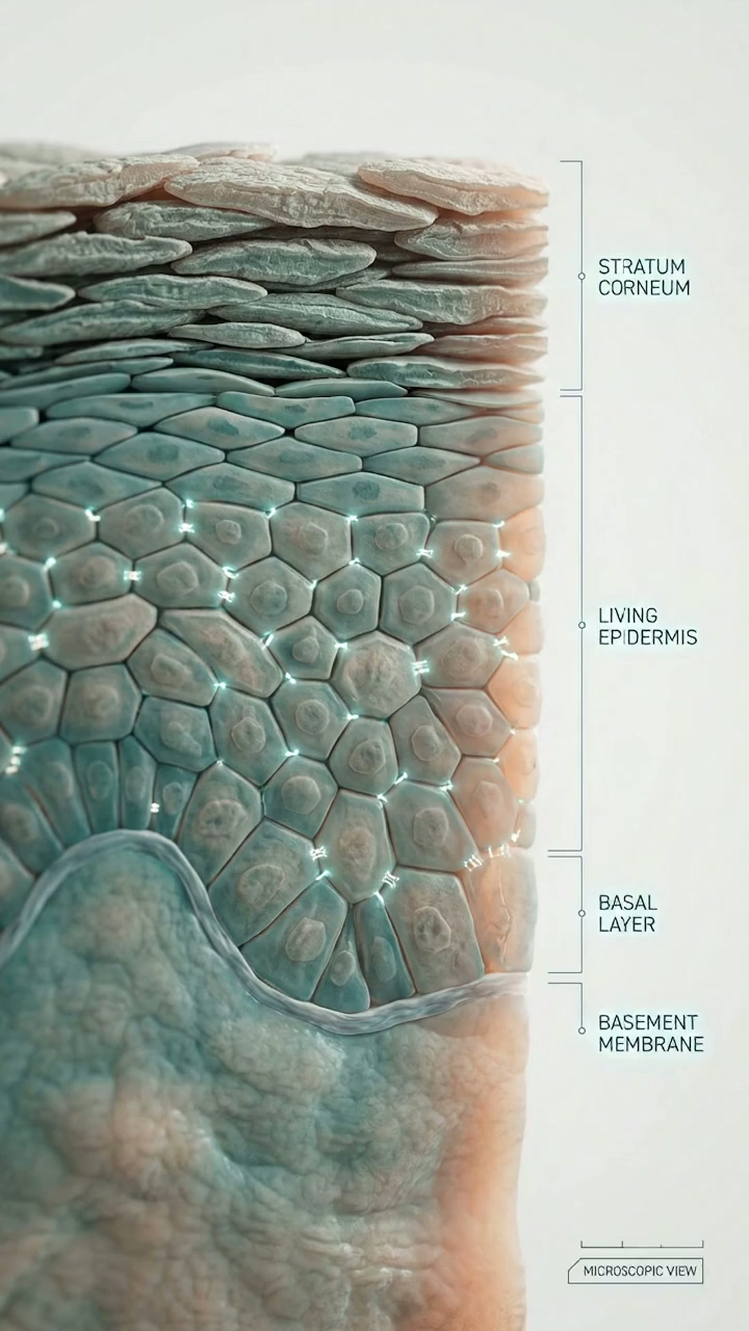
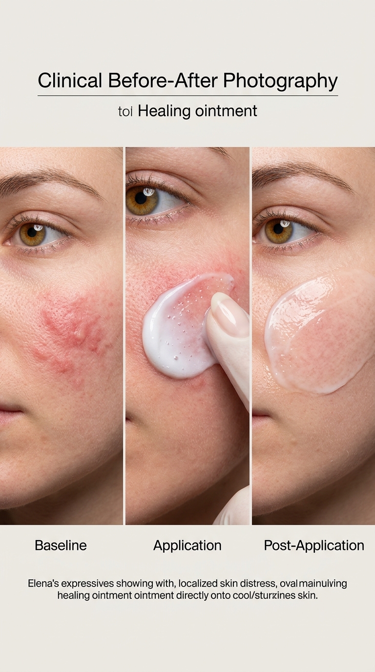
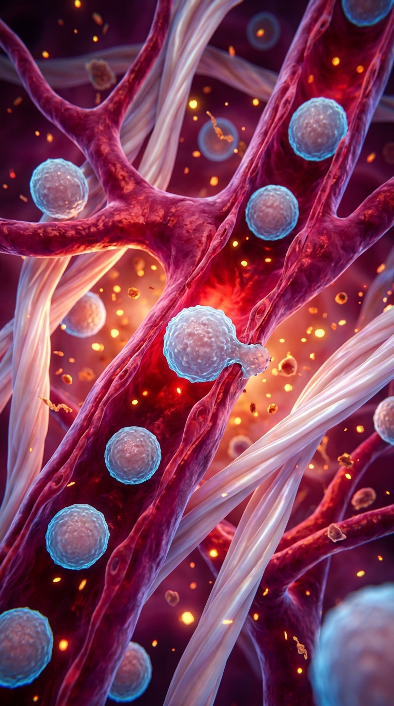





Art style: Clinical Before-After Photography. A microscopic 3D visualization of the dermis layer during an inflammatory response. Large, branching red blood vessels are engorged and pulsing with life. Spherical white blood cells, translucent and textured with small surface bumps, are seen migrating through the vessel walls and into the surrounding interstitial fluid. The fluid is filled with glowing golden cytokines and signaling molecules. The collagen fibers, resembling thick pearlescent ropes, are stretched and distorted by the pressure of fluid accumulation. The lighting is internal and dramatic, with a deep red glow emanating from the vessels and a sharp blue rim light on the immune cells. The atmosphere is chaotic and high-energy. Colors are deep magentas, fiery oranges, and electric blues. The depth of field is narrow, focusing on a single white blood cell as it squeezes through a gap in the capillary wall. Suspended particles of cellular debris and nutrients float like embers in a fire. The texture of the vessel walls is organic and slightly porous. ━━━━━━━━━━━━━━━━━━━━━━━━━━━━━━━━━━━━━━ FINAL STYLE ENFORCEMENT (this is the authoritative rendering directive — it overrides any style wording elsewhere in the prompt): Render this image entirely in "Clinical Before-After Photography" style. Match the lighting grammar, material response, colour science, level of realism / stylisation, line weight, and shading model typical of "Clinical Before-After Photography". Keep the subject, composition, camera angle, lighting direction, and colour palette from the description above — but EVERY surface, skin, fabric, material, and environmental element must be rendered in the "Clinical Before-After Photography" idiom, not in any other medium.
Free to start · Generate videos and images with AI in seconds